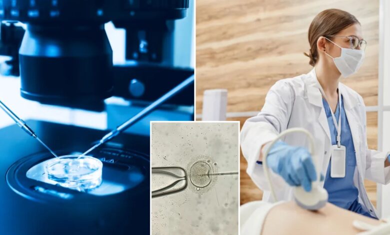
Researchers say they have developed a 3D imaging model of early-stage embryos that may make it easier for women to get pregnant through in vitro fertilization (IVF).
About 2.5% of US births — nearly 92,000 — were from IVF in 2022. When IVF patients create several embryos, it can be difficult to determine which ones have the best chance of resulting in birth.
Now, scientists in China say their 3D model of blastocysts — embryos that are around 5 or 6 days old — can provide previously unknown details about cell features that have been associated with successful pregnancies.
“This study shows that the 3D shape of the blastocyst’s inner cell mass, its position, and how the surrounding cells are arranged can be important indicators of success, which we didn’t know before,” said lead study author Dr. Bo Huang, an embryologist at the Reproductive Medicine Center of Tongji Hospital in China.
In IVF, eggs are collected from a woman and combined with sperm in a lab to create embryos, which are placed into the uterus.
Patients can get their embryos tested for genetic abnormalities before transfer. The success rate is 60% to 65% for genetically healthy embryos. These odds diminish when the woman is older or has uterus conditions that make it difficult for an embryo to implant.
This new study included women younger than 40 with a thick uterine lining and no more than one failed embryo transfer. Using EmbryoScope+, technology that can monitor the development of embryos, researchers took detailed images of 2,141 blastocysts.
They compared their model with fluorescence imaging of the blastocysts and found it was 90% accurate.
“Traditionally, the quality of blastocysts is assessed using 2D methods that lack depth and comprehensive indicators,” Huang explained. “Although some 3D methods exist, they aren’t practical or safe for clinical use. This study bridges that gap by introducing a clinically applicable 3D evaluation method and reveals previously unrecognized … features of blastocysts.”
Huang’s research, published in the journal Human Reproduction, was presented Monday in Amsterdam at the annual meeting of the European Society of Human Reproduction and Embryology (ESHRE).
He said the ultimate goal is to make this technology a standard in IVF, bringing new hope to patients.
Dr. Anis Feki, chair-elect of ESHRE, said Huang’s model “shows great promise,” but further research is needed.
“This method could potentially enhance IVF outcomes, but its clinical application should be approached with careful consideration,” Feki added.